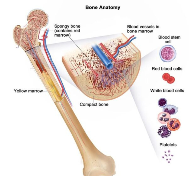
Nuclear Medicine
What is Nuclear Medicine?
Nuclear Medicine (NM) is a specialized medical imaging technique that utilizes small amounts of radioactive isotopes attached to molecules or chemicals, known as radiopharmaceuticals. These radiopharmaceuticals, once administered to the patient, localize within specific organs and organ systems and emit photons, which are then detected by sophisticated equipment called Gamma Cameras. Unlike conventional imaging modalities, such as x-rays, CT scans, or MRIs, Nuclear Medicine provides functional information, including physiological, pathological, molecular, and metabolic details of various tissues and organs within the body.
In addition to diagnosis, Nuclear Medicine also encompasses several therapeutic applications, notably in the treatment of hyperthyroidism, thyroid cancers, advanced neuroendocrine tumors, bone pain resulting from cancer metastasis, and inflammatory joint disorders.
How is the Procedure Conducted?
The procedure typically involves intravenous administration of the radiopharmaceutical. Depending on the type of study, scanning may occur immediately or after a certain period following the administration of the radiotracer. Various images or serial scans may be required in some cases.
What is Nuclear Medicine?
Safety of Nuclear Medicine Procedures
General Instructions
Different Types of Nuclear Medicine Studies

Detects bone cancer, tumors, fractures, or infections. No special preparations are required.
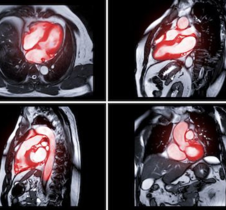
Provides a three-dimensional image of the heart to assess blood flow under stress and resting conditions. Special instructions are provided for this study, and patients may remain in the department for up to five or six hours.
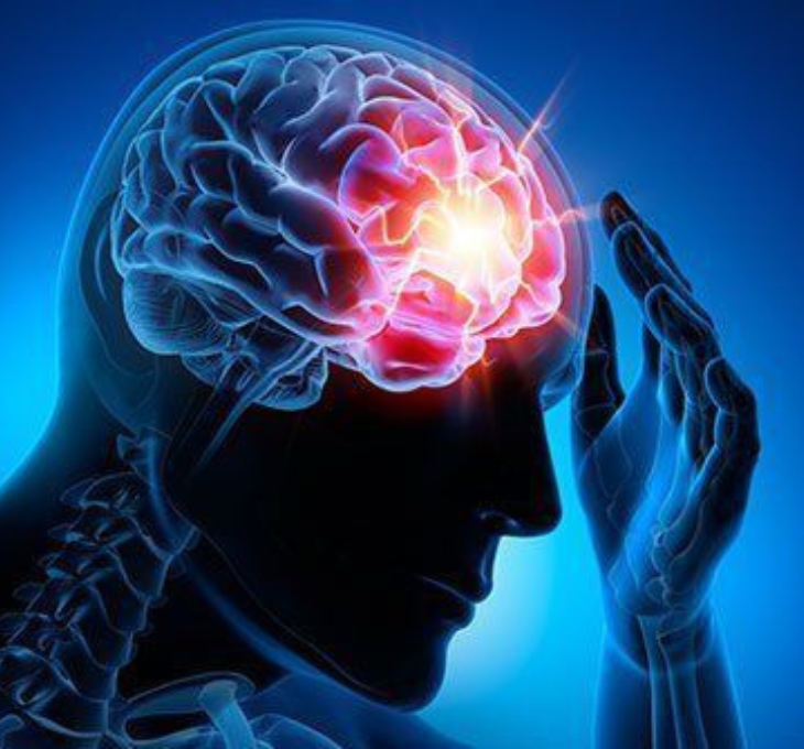
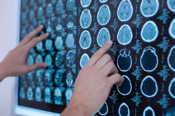
Evaluates blood flow in different brain areas and aids in the diagnosis of dementia, stroke, headaches, or seizures.
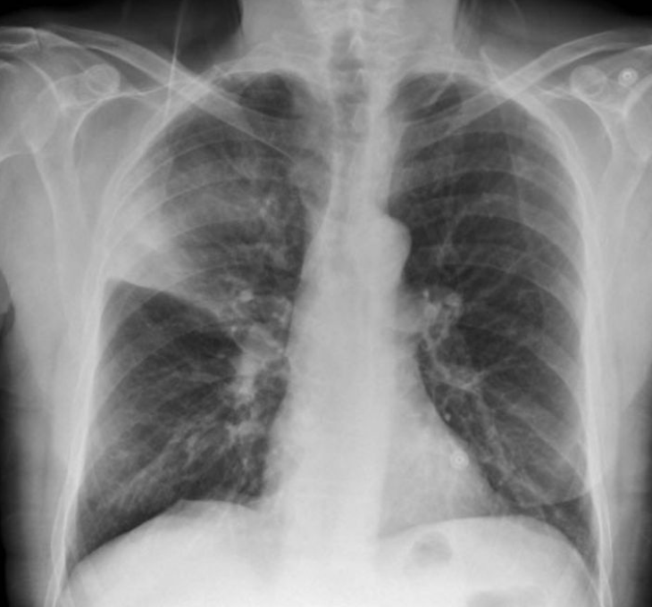
Demonstrates blood supply to the lungs and helps detect blood flow obstructions, such as pulmonary embolism.
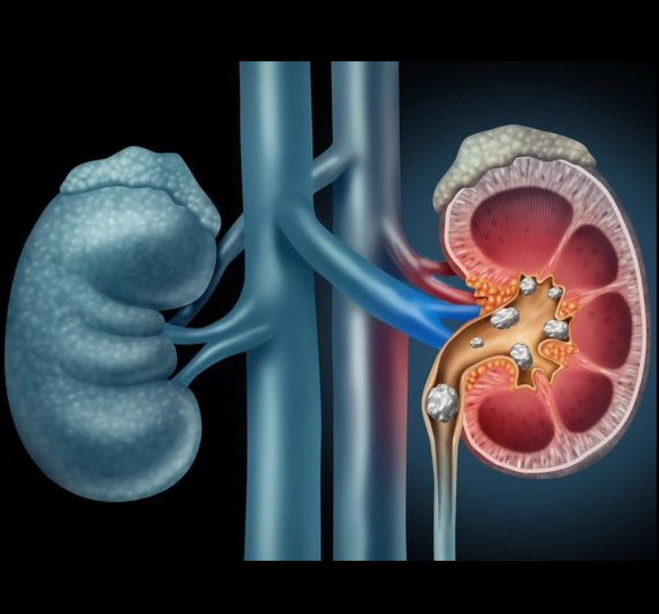
Evaluates kidney function, blood flow to each kidney, and detects urinary flow abnormalities.
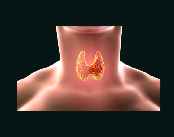
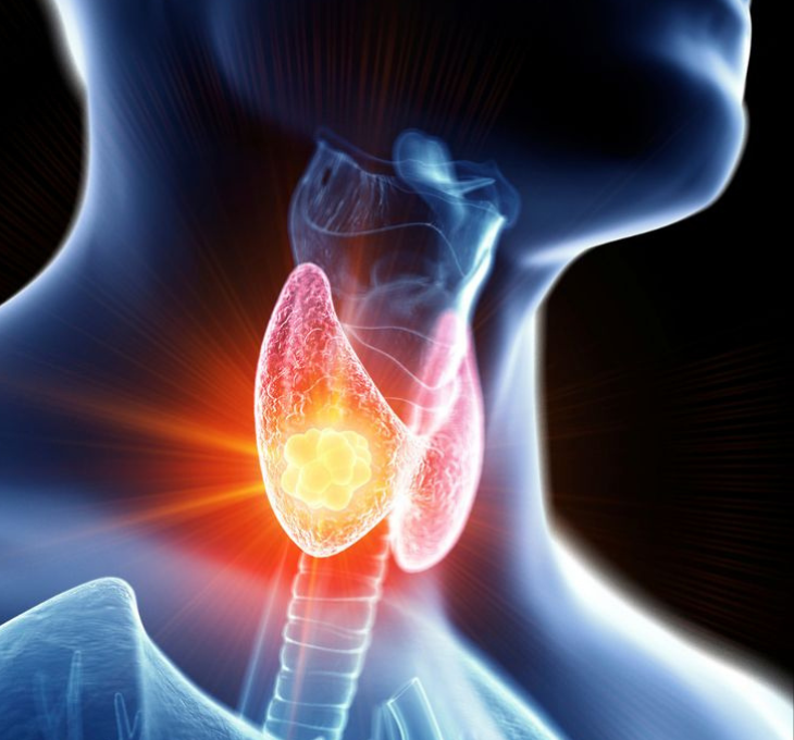
Assesses thyroid gland function, especially hyperactivity.

Performed in thyroid cancer patients after thyroid surgery or during follow-up.
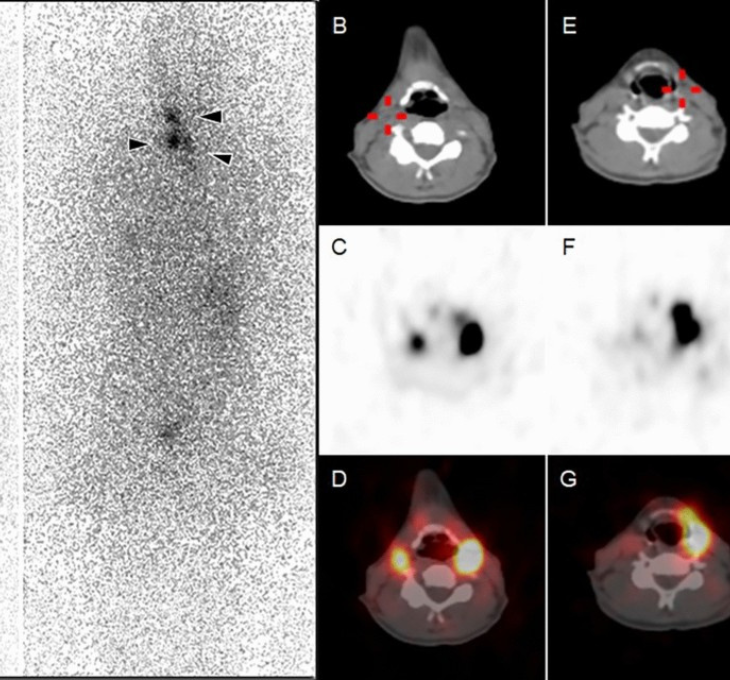
Used to identify parathyroid gland abnormalities causing elevated calcium levels.

Localizes gastrointestinal bleeding sites for appropriate treatment.
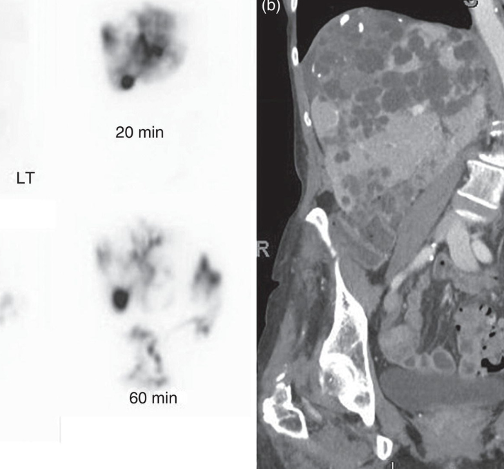
Obtains liver and gallbladder images to diagnose liver dysfunction, gallbladder inflammation, etc.
No WhatsApp Number Found!
WhatsApp us